
-
Find the right food for your petTake this quiz to see which food may be the best for your furry friend.Find the right food for your petTake this quiz to see which food may be the best for your furry friend.Featured products
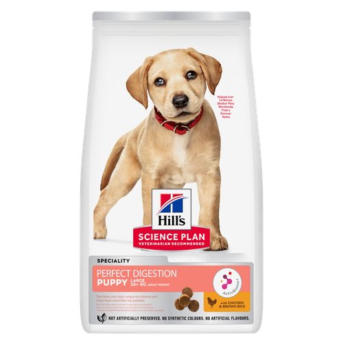 Perfect Digestion Large Breed Puppy Food
Precisely balanced nutrition with Hill's ActivBiome+ prebiotic blend actively contributes to supporting digestive health and overall well-being to help your pet feel their best
Shop Now Small & Mini Mature Adult 7+ Dog Food
Hill's Science Plan Small & Mini Breed Mature Adult Dog Food with Chicken is a complete pet food, specially formulated with ActivBiome+ Multi-Benefit Technology.
Tailored nutrition to support graceful ageing in small dogs. Specially made with a synergistic blend of nutrients for energy & vigor.Shop Now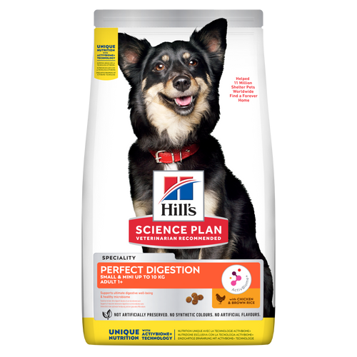 Perfect Digestion Small & Mini Adult Dog Food
Hill's Science Plan Perfect Digestion Small & Mini Breed Adult Dog Food with Chicken & Brown Rice supports ultimate digestive well-being & a healthy microbiome.
Shop NowFeatured products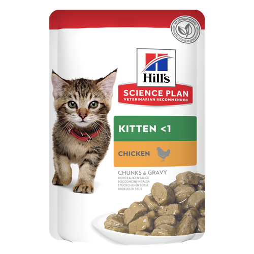 Kitten Food
Tender chicken chunks in gravy for kittens, with omega-3s for healthy eye & brain development and high-quality protein to support muscle growth. With balanced minerals to promote strong bones & teeth.
Shop Now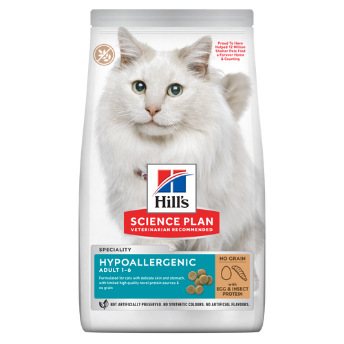 Hypoallergenic Dry Cat Food
HILL'S SCIENCE PLAN Hypoallergenic Adult cat food with egg & insect protein is a complete pet food for adult cat 1–6 years old. It's formulated for cats with delicate skin and stomach, with limited high quality novel protein sources & no grain.
Shop Now Hairball & Perfect Coat Adult Cat Food
Hill's Science Plan HAIRBALL & PERFECT COAT Adult cat food with Chicken is specially formulated to effectively help avoid hairball formation in adult cats while promoting a beautiful coat. Thanks to its mix of essential Omega-6 fatty acids, this food benefits the cat's skin and fur keeping them healthy and shiny. Our Advanced Fibre Technology helps reduce hairballs by naturally promoting their passage through the gut. This food is formulated with high-quality protein for a perfectly balanced, great-tasting recipe.
Shop Now -
Dog
- Dog Tips & Articles
-
Health Category
- Weight
- Food & Environmental Sensitivities
- Urinary
- Digestive
- Joint
- Kidney
-
Life Stage
- Puppy Nutrition
- Adult Nutrition
- Senior Nutrition
Cat- Cat Tips & Articles
-
Health Category
- Weight
- Skin & Food Sensitivities
- Urinary
- Digestive
- Kidney
-
Life Stage
- Kitten Nutrition
- Adult Nutrition
Featured articles Pet Food Storage Tips
Where you store your cat and dog food can make a big difference in the quality and freshness once it is opened. Here are some common questions and recommendations for optimal storage for all of Hill’s dry and canned cat and dog food.
Read More Understanding Your Pet's Microbiome
Learn what a pet's microbiome is, how it contributes to your pet's gut & overall health, and why nutrition is important in maintaining healthy microbiomes.
Read More The Right Diet For Your Pet
Learn what to look for in healthy pet food & nutrition, including ingredients, quality of the manufacturer, your pet's age, and any special needs they have
Read More -


Mast cell tumours in dogs are the most common canine skin tumours, according to the Animal Trust. But what exactly is a mast cell tumour, what are the signs of mast cell tumours in dogs, and can these tumours be treated?
Causes of Mast Cell Tumours in Dogs

Mast cells themselves aren't cancer cells. As the Animal Trust explains, they're a type of white blood cell. They originate in the bone marrow and are found throughout the body's various tissues, where they play a number of roles. These include inflammatory responses, allergic responses, parasite presence responses, and even the breakdown of dead tissues.
Unfortunately, sometimes things go awry. For reasons veterinary scientists don't yet understand, mast cells sometimes mutate and multiply in masses, going against the normal cell life cycle that keeps their numbers in check. This unregulated growth results in a mast cell tumour. These tumours can occur in dogs of all breeds and ages, although the PDSA notes that they are more common in middle-aged dogs and certain breeds, such as boxers or beagles.
Clinical Signs of Mast Cell Tumours in Dogs
The Animal Trust suggests keeping an eye out for the following symptoms of mast cell tumours:
Look for one or more masses anywhere on your dog’s body. They may be visible on (or just below) the skin, muzzle, mouth, or genitals.
Mast cell tumours in dogs usually appear in the form of a lump, but they look like nodules, patches, swellings, or ulcers. They are usually 2-3 cm in diameter.
The mass may ooze or bleed, and there be redness, inflammation, swelling, nodules, or hives around the affected area.
Your dog may have appetite loss, weight loss, gastrointestinal symptoms (e.g. vomiting, diarrhoea) or black/bloody faeces.
When you're petting or examining your dog, look out for any areas of skin that look or feel different. Remember that a cancerous mass may feel firm and tightly adhered to your dog’s body, but it may also be squishy and moveable under the skin. It may grow rapidly in size or remain the same size, but it can appear to recede, too; this doesn’t necessarily mean it’s not dangerous, so it’s important to see your vet to be safe.


Tasty Tips
Diagnosing Mast Cell Tumours in Dogs
Lumpy, bumpy, raised, flat, loose, squishy, firm, big or small — each and every lump on your dog should be evaluated by a veterinarian.
Your vet may very well need to perform a diagnostic test called a fine needle aspiration to collect a small sampling of cells from the lump. Luckily, this test is fast and minimally invasive, and it can usually be done following the initial examination. However, a tumour may not shed many cells during this test, or the sample may not be representative of the whole lesion. So, though this test is a great starting point, your vet may recommend a punch biopsy, as it allows a more comprehensive examination of the mass's architecture. With a biopsy, a microscopic examination of the tissue is sent out to a specialised histopathology lab for a veterinary pathologist to evaluate.
After a mast cell tumour diagnosis, your vet may recommend additional diagnostics such as blood tests, X-rays, CT scans and ultrasounds to determine whether the tumour has spread to other locations (known as metastasis). A fine needle aspirate of a lymph node close to the tumour may also be recommended.

Mast Cell Tumour Treatment
Cancer treatment in dogs is determined by a veterinary oncologist, with input from a veterinary pathologist, who will review the findings of the diagnostic tests and assess the grade, or extent, of the cancer. Mast cell tumours are referred to as “low-grade” or “high-grade”, depending on how advanced they are and whether they have spread. Depending on their findings, the pathologist will then be able to formulate the appropriate treatment plan.
The pathologist may recommend surgery to remove the cancerous mass. The goal is to remove all of the cancerous tissue, along with a border of healthy tissue around the edges to be certain that no cancer cells are left behind. This is called achieving “clean margins”. The pathologist will examine the surgically removed tissue afterwards to make sure this has been achieved, although it is not always possible.
Surgery alone may be enough to treat a mast cell tumour, but in some cases it might be done alongside chemotherapy or radiotherapy. The Animal Trust says that, in unsuccessful or advanced cases, oral medications might be recommended to reduce the size or slow the growth of the tumour. Steroids may also be given to reduce inflammatory effects caused by the tumour.
While these approaches are often strongly recommended, especially at more advanced stages, keep in mind that you can also choose to focus on gentler approaches or comfort care at any stage. Your vet and/or veterinary oncologist will be happy to discuss these options with you.
Life Expectancy for Dogs With Mast Cell Tumours
It is difficult to predict the life expectancy for dogs who develop mast cell tumours, as it varies depending on many factors. If the tumour is limited to the skin with no evidence of metastasis, and it is removed with clean margins, the prognosis can be quite positive. However, for higher-grade tumours, or those in locations such as the mouth or genitals, the prognosis may not be as strong.
The good news is that early diagnosis and treatment improve the prognosis of mast cell tumours, so it’s important to regularly examine your dog at home. This can be done simply with observational petting and intentional at-home exams. Remember to schedule a veterinary visit should you discover any new masses on your dog.


Dr. Laci Schaible is a small animal veterinarian, veterinary journalist, and a thought leader in the industry. She received her Doctor of Veterinary Medicine from Texas A&M University and her Masters in Legal Studies from Wake Forest University.
Related products
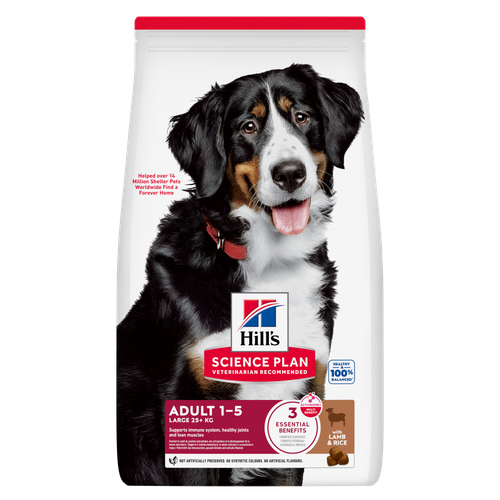
Hill's Science Plan Large Breed Adult Dog Food with Lamb & Rice is a complete pet food, specially formulated with ActivBiome+ Multi-Benefit Technology.
This food is specifically designed to fuel the energy needs of large breed dogs during the prime of their life.

Hill's Science Plan Perfect Digestion Small & Mini Breed Adult Dog Food with Chicken & Brown Rice supports ultimate digestive well-being & a healthy microbiome.

Precisely balanced nutrition with Hill's ActivBiome+ prebiotic blend actively contributes to supporting digestive health and overall well-being to help your pet feel their best

Hill's Science Plan Small & Mini Breed Mature Adult Dog Food with Chicken is a complete pet food, specially formulated with ActivBiome+ Multi-Benefit Technology.
Tailored nutrition to support graceful ageing in small dogs. Specially made with a synergistic blend of nutrients for energy & vigor.
Related articles

Dog obesity is a significant problem - learn more about helping your dog become trimmer and healthier through improved nutrition.

Discover how the field of dog science is giving us more and more insights into the inner workings of our furry best friends.

Learn about snake bites on dogs, including clinical symptoms to look for, what to do if you think your dog was bitten, and treatment & prevention options.

Discover the causes, signs, and treatments of kidney disease in dogs and find methods of supporting your dog's kidney health. Learn more at Hill's Pet South Africa.

Put your dog on a diet without them knowing
Our low calorie formula helps you control your dog's weight. It's packed with high-quality protein for building lean muscles, and made with purposeful ingredients for a flavorful, nutritious meal. Clinically proven antioxidants, Vitamin C+E, help promote a healthy immune system.

